Eat, Sleep, Fight, Repeat: Macrophage Cells at Work
14th June 2022 - Last modified 19th October 2023
20 years of Alto. 20 years of science. #6
By Martina Neville PhD, Senior Manager, SEO and Content Marketing
As part of Alto Marketing’s 20 year celebrations, we’re looking back at some of the most important advances in science over this time in our blog series “20 years of Alto. 20 years of science.” We gave each of the scientists in the Alto team the chance to write about an area they love or that they’ve worked on during their research careers. Here, Martina Neville, Senior Manager, SEO & Content introduces the workhorses of the immune system, macrophages. From COVID-19 to cell therapy, discover the most influential macrophage discoveries of the past 20 years.
The macrophage. An immune shapeshifter. A cellular paradox. The underdog of the immune response…
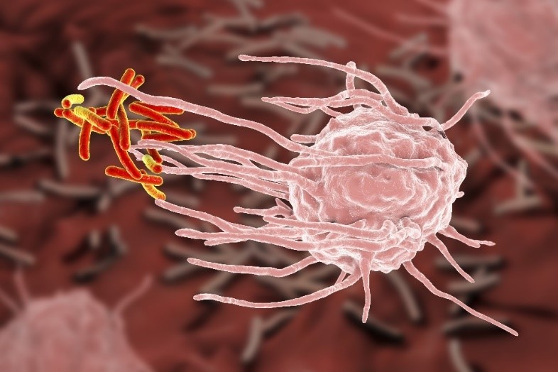
Most of us familiar with macrophage cells will recognise them for their ability to phagocytose (eat) invading material and cellular debris.
But in addition to their well-established role as eating machines, they also do much, much more! In fact, over the last two decades, we’ve only just scratched at the surface of their abilities. From infection and tissue regeneration through to cancer and autoimmunity, macrophages play an all-encompassing role in a plethora of immune-related scenarios.
But why am I, a senior account manager in life science and healthcare marketing, writing a blog about macrophages?
Because first and foremost I am a scientist. And it really all comes down to my one obsession – immunology. In fact, I’ve spent most of my adult life fascinated and in awe of the intricacies of immune cells. Notably, their ability to adapt to the harshest of conditions to protect us but also how they can turn on us at a moment’s notice – or as I like to call it, ‘immune friendly fire’.
But it was during my PhD that I developed a true passion for the humble macrophage. And in my opinion, macrophages are enormously underestimated and rarely given the attention they deserve.
In this blog, we’ll be taking you on an unforgettable journey through some of the most prominent macrophage advances of the past 20 years. By the end, I hope that not only will you be au fait with the macrophage but also be persuaded that there’s still much to be gained from macrophage research.
An identity crisis? Or an immune chameleon?
“Can you remember who you were, before the world told you who you should be?” – Charles Bukowski”
Like all good stories, this starts at the very beginning and tells a familiar tale of environment influencing behaviour.
For many years we thought that macrophages started their life as monocytes within the bone marrow, before being released into the blood, where they circulated, migrating to specific tissue sites for specialised differentiation. Now, while that may be true in a few cases such as the gut, dermis and heart, fate mapping studies have since revealed that most tissue resident macrophages are in fact, formed from embryonic precursors1.
Polarized into a number of phenotypes, it will come as no surprise to those familiar with immunology to hear that there are two distinct activated sub-types that live at opposite ends of the ‘spectrum’: the pro-inflammatory, immunogenic and antitumor M1 and the immunosuppressive, tolerogenic and protumour M22.
But our story doesn’t end there. Macrophages also display a high degree of functional plasticity, that is, they may transition from one state to another at any one time depending on the environment in which they exist. In fact, when it comes to macrophage behaviour, it’s the surrounding environment and associated milieu of cytokines/chemokines that are most influential.
Tumour-associated macrophages (TAMs) – A special kind of macrophage
The complexities of the tumour microenvironment (TME) are colossal, with a paradoxical landscape of cells and cytokines that are pro-inflammatory, immune-suppressive and somewhere in-between at any one time; the proportion of which is determined by the intricacies of the environment. A self-serving environment, the TME remodels its surroundings to increase its chances of a more favourable outcome.
But, where exactly do macrophages fit into this?
In the early stages of tumour progression – and much like MI5 agents – M1 tumour-associated macrophages (TAMs) survey the surrounding area and identify their targets. Unlike MI5 agents, they then kill their targets by scavenging, ingesting, and destroying them – unleashing a barrage of cytotoxic molecules such as nitric oxide (NO) and reactive oxygen species (ROS) in the process.
But it’s when tumour cells begin to dominate the local area that macrophages are forced to adapt, influenced by the expression of markers on the tumour cell surface, such as CD47 and PD-L1, and by surrounding soluble factors. Essentially, macrophages are ‘hypnotised’ and lulled into a relaxed state where they take on a pro-tumour M2 phenotype.
When it comes to TAM sub-types, one could be forgiven for thinking that the two states are mutually exclusive, however, an increasing number of studies allude to the possibility of single-cell bifunctionality.
Breaking all of the supposed ‘rules’ of cell polarization, it turns out that in addition to typical M1/M2 phenotypes, they can simultaneously express M1 and M2 gene signatures 3 in readiness for external stimuli.
Tissue remodelling and regeneration: macrophage cells at work
To understand how macrophages are involved in tissue regeneration we must first understand the basics of non-infectious and infectious injury.
Non-infectious injury
In the absence of a pathogen, necrosis leads to the death of affected cells causing the rupture of cell membranes. As a result, a number of intracellular components known as Damage-Associated Molecular Patterns (DAMPs) are released into the local environment. The interaction of DAMPs with DAMP-sensing receptors triggers sterile inflammation.
Infectious injury
Characterised by the presence of a pathogen, infected injuries lead to the release of Pathogen-Associated Molecular Patterns (PAMPs) as well as DAMPs. Following recognition by Pattern Recognition Receptors (PRRs) on or within immune cells, acute inflammation occurs.
Repair/Remodelling
When injury occurs, much like paramedics first to the scene, M1 macrophages flock to the site of injury to phagocytose pathogens and debris from dying cells, while also releasing chemotactic agents to recruit more immune cells. In the resolution phase of inflammation, M2 macrophages secrete a plethora of factors. Each of these factors plays a key role in the promotion of angiogenesis, and cellular proliferation – to initiate fibrosis and remodel the extracellular matrix.
As with anything in life, it’s all about balance.
And when macrophages go rogue, repair and regeneration can become a huge problem. In renal disease, M2 macrophages, transition to myofibroblasts to promote renal fibrosis, a major driver of progression to end-stage renal disease4. A similar pattern has been observed in several other tissue types including cardiac5, brain6, and lung7.
A double-edged sword in COVID-19
Unsurprisingly, the COVID-19 pandemic has caused a huge outbreak of immunology research. And the macrophage is no newcomer to the world of coronavirus diseases. In fact, since the first outbreak of SARS-CoV in 2002 – 2003, scientists have suspected that macrophages are a key player in SARS disease progression8.
It’s only now, as we tiptoe gingerly out of the pandemic, that we begin to truly comprehend the significance of macrophages in SARS-CoV pathogenesis.
While several studies have suggested that monocyte-derived interstitial macrophages are prevalent in patients with late-stage COVID-19, in some cases resident alveolar macrophages appear to be severely depleted9,10. This interesting finding may indicate that specific types of macrophages are responsible for the severe lung fibrosis seen in patients with later-stage COVID-19.
But what exactly is happening to the macrophage during COVID-19 infection?
Truth be told, it’s likely to be a complex process, far too substantial to be covered in this short blog. However, interestingly, the SARS-CoV-2 spike protein (S-protein) appears to act as a viral PAMP, initiating the assembly of an immunomodulatory complex in COVID-19 patient-derived macrophages11. But more than that, inflammasome activation is dependent on prior priming of monocytes suggesting that SARS-CoV2 S-protein induces a state of innate immunological memory.
Macrophage engineering
While every other immune cell was being engineered, it appeared that macrophages were left in the dark. However, it seems that scientists are now harnessing the humble macrophage to target tumours. The phagocytic nature of some macrophages is an attractive feature to scientists and recent studies indicate that they can be engineered to deliver drug nanoparticles12.
In addition, companies such as Carisma Therapeutics and Thunder Biotech are in the process of developing Chimeric Antigen Receptor (CAR) macrophages. In fact, preliminary results at Penn Medicine, in the very first in-human cell therapy trial with CAR macrophages, indicate that the therapy is safe to use.
Jack-of-all-trades and master of all
Hopefully, by now I’ve managed to convince you that the macrophage is not a cell to be ignored. In true court-case style, I’ve sworn to tell the whole truth, I’ve taken the stand and I’ve given ample evidence as to why macrophages should be taken more seriously.
Now, it’s down to you, the jury, to decide whether or not the evidence speaks for itself.
About me:
I don’t have an exciting story to tell about how I became a researcher. But, I love research and it still plays a large part of my everyday life, albeit not in the lab. During my undergraduate degree, I was lucky enough to be in the presence of two very passionate lecturers, who subsequently went on to be my MRes and PhD supervisors. They played a large part in what was to be my research pathway. I remember vividly sitting in those lectures and being in awe of the immune system and in particular, immunoparasitology (I’ll talk more about this in a later blog).
It’s well known that parasitic infections, particularly those affecting the gastrointestinal tract, have declined drastically in the western world over the past several decades. And it’s this trend, along with the increase in autoimmune disease, that still fascinates me to this day.
It’s perhaps not surprising to hear that my PhD research primarily focused on characterising the macrophage response to parasite-derived homologues of an immunomodulatory protein.
References
1. Hoeffel G, Ginhoux F. Ontogeny of tissue-resident macrophages. Frontiers in Immunology. 2015;6(SEP):486. doi:10.3389/FIMMU.2015.00486/BIBTEX
2. Chávez-Galán L, Olleros ML, Vesin D, Garcia I. Much more than M1 and M2 macrophages, there are also CD169+ and TCR+ macrophages. Frontiers in Immunology. 2015;6(MAY):263. doi:10.3389/FIMMU.2015.00263/BIBTEX
3. Garrido-Martin EM, Mellows TWP, Clarke J, et al. M1hot tumor-associated macrophages boost tissue-resident memory T cells infiltration and survival in human lung cancer. Journal for ImmunoTherapy of Cancer. 2020;8(2):e000778. doi:10.1136/JITC-2020-000778
4. Tang PMK, Nikolic-Paterson DJ, Lan HY. Macrophages: versatile players in renal inflammation and fibrosis. Nature Reviews Nephrology 2019 15:3. 2019;15(3):144-158. doi:10.1038/s41581-019-0110-2
5. Wynn TA, Vannella KM. Macrophages in tissue repair, regeneration, and fibrosis. Immunity. 2016;44(3):450. doi:10.1016/J.IMMUNI.2016.02.015
6. Aldrich A, Kielian T. Central Nervous System Fibrosis Is Associated with Fibrocyte-Like Infiltrates. The American Journal of Pathology. 2011;179(6):2952. doi:10.1016/J.AJPATH.2011.08.036
7. Ogawa T, Shichino S, Ueha S, Matsushima K. Macrophages in lung fibrosis. International Immunology. 2021;33(12):665-671. doi:10.1093/INTIMM/DXAB040
8. Perlman S, Dandekar AA. Immunopathogenesis of coronavirus infections: implications for SARS. Nature Reviews Immunology 2005 5:12. 2005;5(12):917-927. doi:10.1038/nri1732
9. Rendeiro AF, Ravichandran H, Bram Y, et al. The spatial landscape of lung pathology during COVID-19 progression. Nature 2021 593:7860. 2021;593(7860):564-569. doi:10.1038/s41586-021-03475-6
10. Chen ST, Park MD, Valle DM del, et al. Shift of lung macrophage composition is associated with COVID-19 disease severity and recovery. bioRxiv. Published online January 12, 2022. doi:10.1101/2022.01.11.475918
11. Theobald SJ, Simonis A, Georgomanolis T, et al. Long-lived macrophage reprogramming drives spike protein-mediated inflammasome activation in COVID-19. doi:10.15252/emmm.202114150
12. Ding X, Sun X, Cai H, et al. Engineering Macrophages via Nanotechnology and Genetic Manipulation for Cancer Therapy. Frontiers in Oncology. 2021;11. doi:10.3389/FONC.2021.786913